Medical Imaging and Acupuncture for Pain Control
Medical imaging technology allows us to see the effects of acupuncture as a treatment is occurring. There is a significant body of research using PET scans, MRI’s and SPECT images? that show the effectiveness of acupuncture. For example, PET scans have shown the link between distal acupuncture points and correlating brain centers–links long ago established in Chinese Medicine. Further, imaging has shown the homeostatic effects of acupuncture, or its ability to restore the brain to more normal functioning (1).
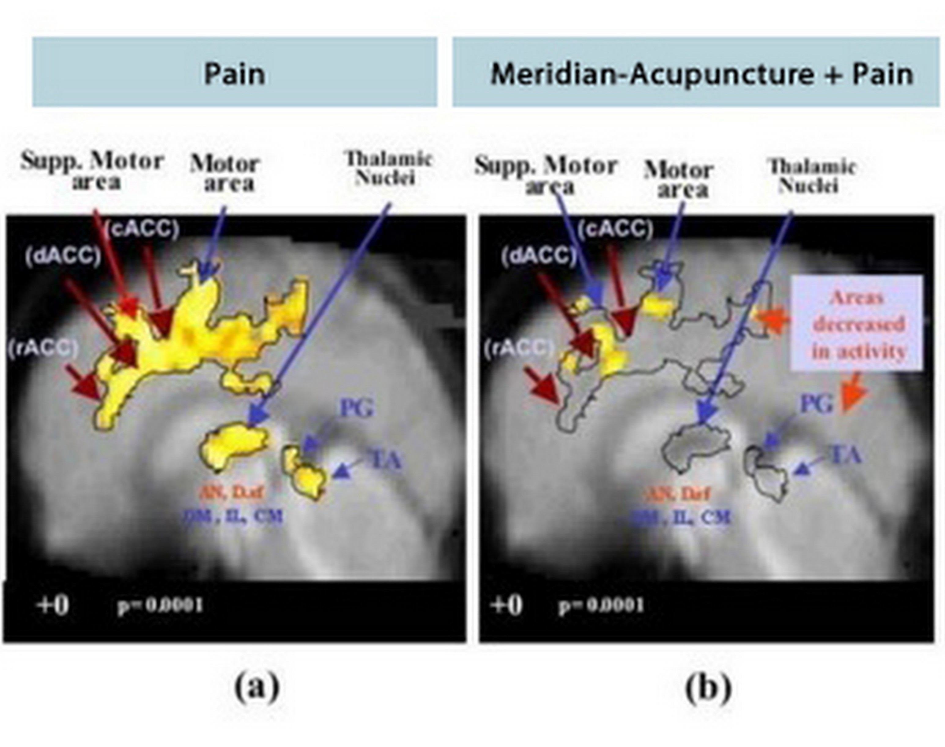
PETscan and Pain Control with Acupuncture
People seeking pain control have often turned to acupuncture for help with great success. Medical imaging shows us why. Acupuncture activates regions of the brain that are active during both acute and chronic pain (2).
The PET scan image below shows areas active during pain on the left and the same areas during acupuncture on the right (3). The decrease in yellow indicates the pain relief of an acupuncture treatment.
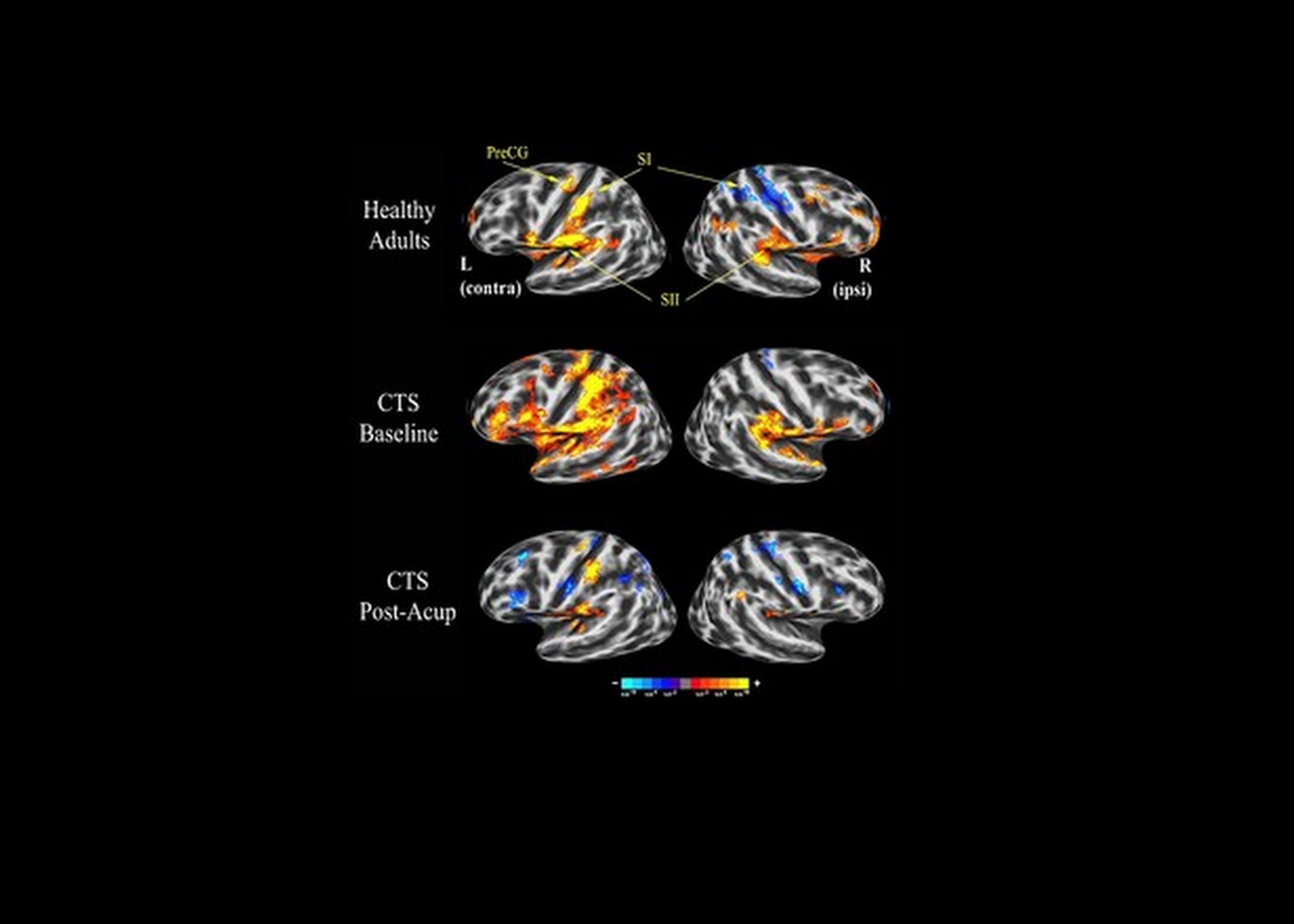
 MRI Images and Pain Control with Acupuncture
Another example below uses MRI images to show the effects of acupuncture treatment for carpal tunnel syndrome. We can see the brain of a healthy subject in the top row. The middle two images show the pain of a patient with carpal tunnel syndrome indicated by red and yellow areas. The bottom two images show the brain of the patient with carpal tunnel syndrome after acupuncture (4).
SPECT Imaging and Pain Control with Acupuncture
A study done by Abass Alavi, M.D. used SPECT imaging to show acupuncture effects in the brain. He viewed the brains of four people with pain and compared these to five pain-free people. Dr. Alavi found that after acupuncture needles were inserted, all of the subjects had increased blood flow to the thalamus and basal ganglia. These regions are involved in the processing of pain signals.
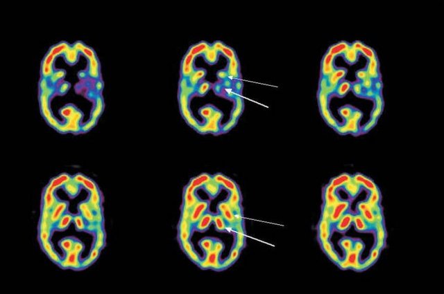
The top row of images show the initial activity of the thalamus (thick arrow) and the basal ganglia (thin arrow) in a patient with pain. The activity is unequal with less on the right. After acupuncture, functioning is normalized with right and left having greater and equal activity. The activity changes indicate lessened pain and the brain restored to more normal functioning (5).
Jeffrey Russell is an acupuncturist and practitioner of Chinese herbalism in Louisville. You can reach him at his clinic, Abacus Chinese Medicine, at 502 299-8900.
![]()
Works Cited
-
Zang-He, C., Olsen, T.D., Alimi, D., Niemtzow, R.C. Acupuncture: The Search for Biologic Evidence with Functional Magnetic Resonance Imaging and Positron Emission Tomography Techniques. J. Alt Compl. Med. 2002 8(4) 399
-
Biella G1, Sotgiu ML, Pellegata G, Paulesu E, Castiglioni I, Fazio F.Neuroimage. Acupuncture produces central activations in pain regions. 2001 Jul;14(1 Pt 1):60-6.
-
Source: Dr. Zang Hee Cho, et al.
-
Source: Vitaly Napadow
-
Alavi A, Lee L, Farrar J, Lee B, Lariccia P, Newberg A. Cerebral Blood Flow Effects of Pain and Acupuncture: A Preliminary Single-Photon Emission Computed Tomography Imaging Study. Journal of Neuroimaging. 02/2005; 15(1):43-9.